PLATE XIV.
VESSELS OF UMBILICAL CORD.

PLATE XIV. Vessels of umbilical cord.
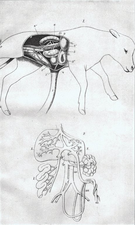
Fig. 1. Fetal calf with a portion of the wall of the abdominal cavity of the right side and the stomach and intestines removed to illustrate the nature of the umbilical or navel cord. It consists of a tube (1-1') into which pass the two umbilical arteries (3) carrying blood to the placenta in the uterus or womb and the umbilical vein (4) bringing the blood back and carrying it into the liver. The cord also contains the urachus (2') which carries urine from the bladder (2) through the cord. These vessels are all obliterated at birth. 5, liver; 5', lobe of same, known as the lobus Spigelii; 5'', gall bladder; 6, right kidney; 6', left kidney; 6'', ureters, or the tubes conducting the urine from the kidneys to the bladder; 7, rectum, where it has been severed in removing the intestines; 8, uterus of the fetus, cut off at the anterior extremity; 9, aorta; 10, posterior vena cava. (From Fürstenberg-Leisering, Anatomie und Physiologie des Rindes.)
Fig. 2. Blood vessels passing through the umbilical cord in a human fetus. (From Quain's Anatomy, vol. 2.) L, liver; K, kidney; I, intestines; U C, umbilical cord; Ua, umbilical arteries. The posterior aorta coming from the heart passes backward and gives rise to the internal iliac arteries, and of these the umbilical arteries are branches. Uv, umbilical vein; this joins the portal vein, passes onward to the liver, breaks up into smaller vessels, which reunite in the hepatic vein; this empties into the posterior vena cava, which carries the blood back to the heart.